70以上 e coli cell wall structure 269097-E.coli cell membrane composition
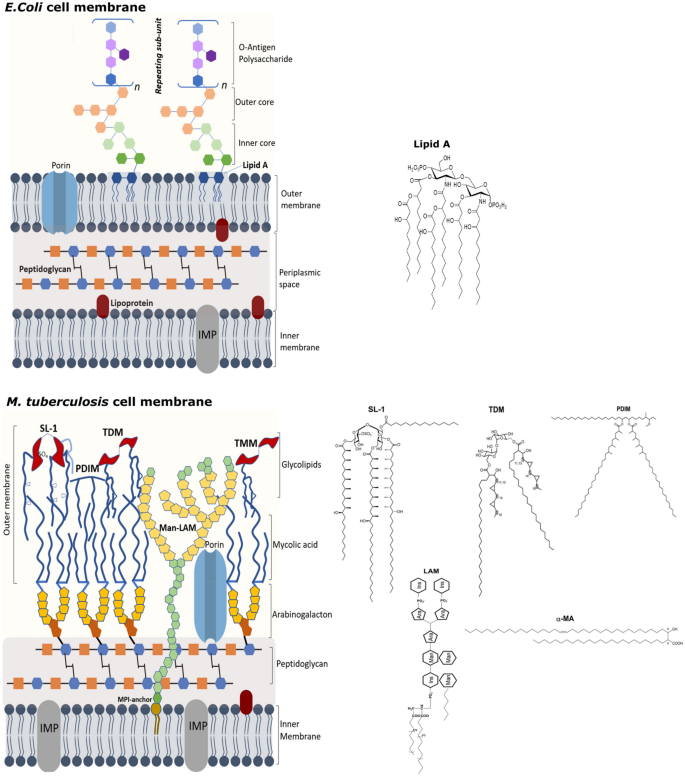
Therefore, ProP also plays an important role in the structure of membrane wall in E coli The concentration of E coli cell bipolar permeability transporter ProP increases with the increase of cardiolipin content and depends on the structure of the cytoplasmic carboxyl terminal domain of the transporter 42Structure And Function Of A Ecoli Bacteria E coli is a rodshaped bacterium The peptidoglycan cell wall is thin and multilayered There is a thin peptidoglycan layer placed between the inner cytoplasmic membrane, and the outer membrane The outer membrane is surrounded by the capsule layer which is composed of polysaccharides The flagellum of E coli consists of three distinct parts the filament, the hook and the basal body all of which are made up of different types of proteinsCells are typically rodshaped, and are about μm long and 025–10 μm in diameter, with a cell volume of 06–07 μm 3 E coli stains Gramnegative because its cell wall is composed of a thin peptidoglycan layer and an outer membrane During the staining process, E coli picks up the color of the counterstain safranin and stains pink

Diagram Demonstrating Of The Cell Wall Structure Of A Grampositive Download Scientific Diagram
E.coli cell membrane composition
E.coli cell membrane composition-Cell Morphology of Ecoli Structure of Cell Wall of Ecoli Antigenic Structure About the Organism Ecoli is considered a gramnegative bacterium based on the gram staining procedure developed by Hans Christian Gram It has a thin peptidoglycan layer and an outer thin layer of lipid due to which it cannot retain the crystal violet stainAlthough the E coli cell wall normally maintains a cylindrical shape during exponential growth , the cell shape can be altered either genetically or environmentally E coli mutants lacking the high molecularweight PBP2, a transpeptidase, swell up to resemble spheroplasts ( 16 ), while cells lacking the low molecularweight PBPs 5 and 7 are often branched with 3 or more poles ( 17 , 18 )


Peptidoglycan Remodeling Enables Escherichia Coli To Survive Severe Outer Membrane Assembly Defect Mbio
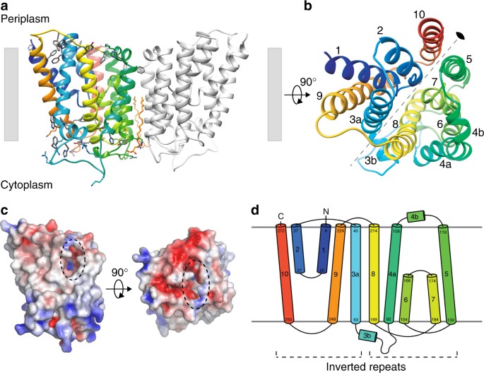
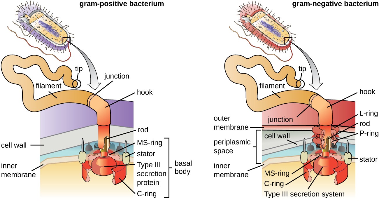
It is different in structure than that of the filament Hook connects filament to the motor portion of the flagellum called a basal body Structure of Bacterial flagella The basal body is anchored in the cytoplasmic membrane and cell wall There are presence of rings that are surrounded by a pair of proteins called MotTogether with the previously reported structure of T africanus MurJ, the structure and saturating mutagenesis of E coli MurJ provides a framework for the design of future experiments to investigate unresolved aspects of MurJ function These include identification of the energetic factor(s) driving lipid II flipping and understanding how MurJ and other proteins in the PG synthesis machinery work in concert to assemble the cell wallFimH, PapG and GafD are twodomain structures connected by a flexible linker, and the Nterminal adhesin domains have an elongated betabarrel jelly roll fold that contains the receptorbinding groove The adhesin domains differ in disulfide patterns, in size and location of the ligandbinding groove, as well as in mechanism of receptor binding
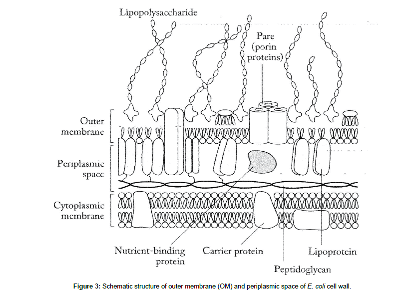
Acid fastness is a physical property of bacteria, which rely on the structure of the bacterial cell wall Typically, the cell wall of bacteria is made up of proteins, carbohydrates, and lipids Acid fast bacteria comprise a thin layer of peptidoglycans The mycolic acid is a long chain of fatty acids, attached to the peptidoglycansHow the mechanical properties of the cell wall are determined by the molecular features and the spatial arrangement of the relatively thin strands in the larger cellularscale structure is not known To examine this issue, we have developed and simulated atomicscale models of Escherichia coli cell walls in a disordered circumferential arrangement The cellwall models are found to possess an anisotropic elasticity, as known experimentally, arising from the orthogonal orientation of theE coli is a Gramnegative rodshaped bacteria, which possesses adhesive fimbriae and a cell wall that consists of an outer membrane containing lipopolysaccharides, a periplasmic space with a peptidoglycan layer, and an inner, cytoplasmic membrane Some strains are piliated and capable of accepting and transferring plasmid to and from other bacteria
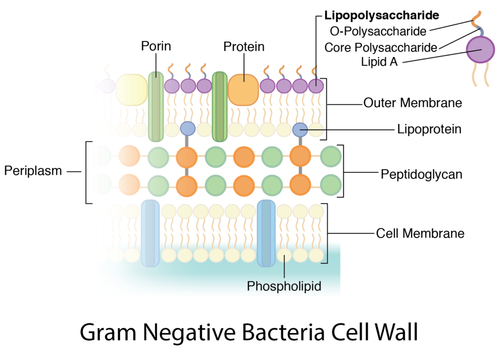
In the Gramnegative Bacteria the cell wall is relatively thin (10 nanometers) and is composed of a single layer of peptidoglycan surrounded by an outer membrane P eptidoglycan structure and arrangement in E coli is representative of all Enterobacteriaceae, as well as many other Gramnegative bacteriaGramnegative bacteria such as Escherichia coli are surrounded by an outer membrane, which encloses a peptidoglycan layer Even if thinner than in many Grampositive bacteria, the peptidoglycan in E coli allows cells to withstand turgor pressure in hypotonic medium In hypertonic medium, E coli treated with a cell wall synthesis inhibitor such as penicillin G form walldeficient cellsOne of the most popular pathogenic bacteria is Escherichia Coli or commonly called E coliMany people know that E coli can cause diarrhea if they ate foods or drinks that are contaminated with E coliActually, there are many types of E coli that can cause different types and level of diarrhea This assignment is going to discuss on the E coli itself, its structure, ecology and mode of
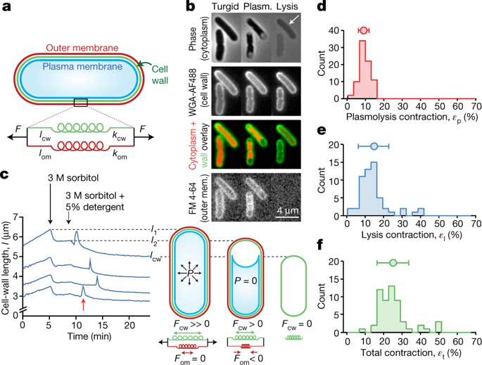


The Outer Membrane Is An Essential Load Bearing Element In Gram Negative Bacteria Nature X Mol
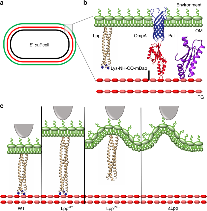


Lipoprotein Lpp Regulates The Mechanical Properties Of The E Coli Cell Envelope Nature Communications
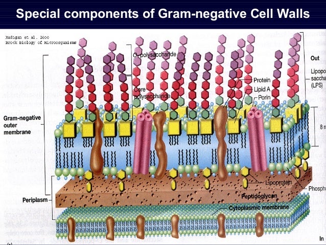
It is in fact an integral compartment of the gramnegative cell wall Together the plasma membrane and the cell wall (outer membrane, peptidoglycan layer, and periplasm) constitute the gramnegative envelope (5, 9) Our entire perception of grampositive and gramnegative walls ultimately relies on the response of bacteria to Gram stainingThe structure of peptidoglycans of Escherichia coli and Bacillus subtilis is similar except for a few minor modifications, but murein (cell wall) structures are extremely different because the major cell wall constituents, anionic polymers, are not attached to peptidoglycans of E coli but are attached to those of B subtilis Thickness of the cell walls in B subtilis and the presence of an outer membrane in E coli are other important differences in the cell wallE coli are rodshaped bacterium that has an outer membrane consisting of lipopolysaccharides, inner cytoplasmic membrane, peptidoglycan layer, and an inner, cytoplasmic membrane Cell wall this structure is made of a thick layer of protein and sugar that prevents the cell from bursting Plasma membrane this structure is made of lipids and proteins and is used to control the movement of molecules in and out the cell


Nlpd Links Cell Wall Remodeling And Outer Membrane Invagination During Cytokinesis In Escherichia Coli


Peptidoglycan Remodeling Enables Escherichia Coli To Survive Severe Outer Membrane Assembly Defect Mbio
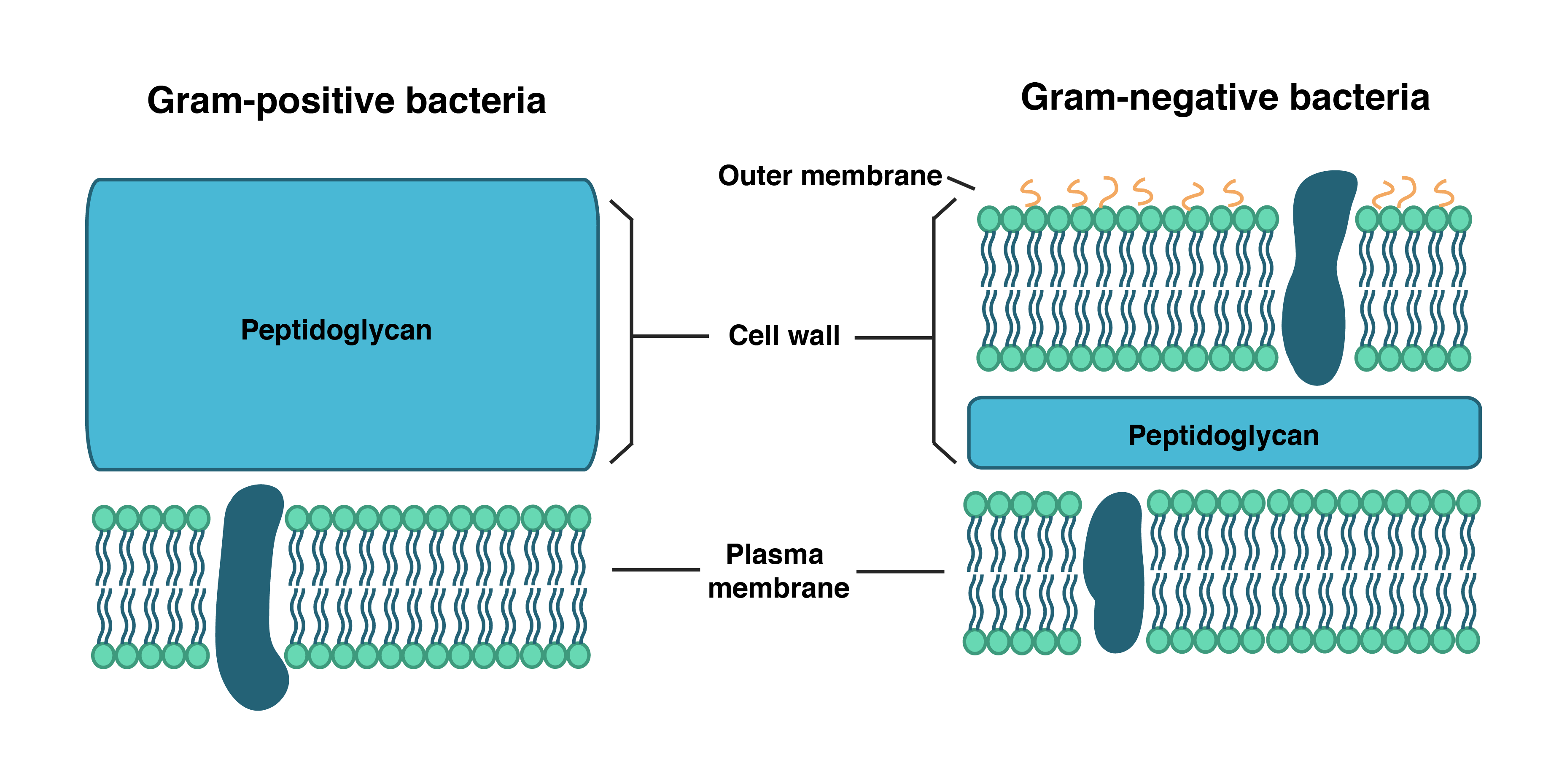
The cell wall is directly exposed to the extracellular milieu in Grampositive bacteria, but is shielded in Escherichia coli and other Gramnegative species by a highly selective permeability barrier formed by the outer membrane (OM)The end result of Escherichia coli morphogenesis is a cylindrical tube with hemispherical caps How does this shape come about?Bacterial Cell wall Structure, Composition and Types Cell wall is an important structure of a bacteria It give shape,rigidity and support to the cell On the basis of cell wall composition, bacteria are classified into two major group ie Gram Positive and gram negative Types of cell wall 1 Gram positive cell wall
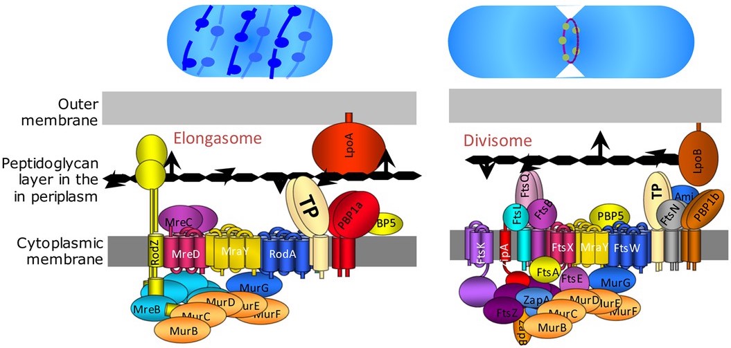


Divisome Wikipedia
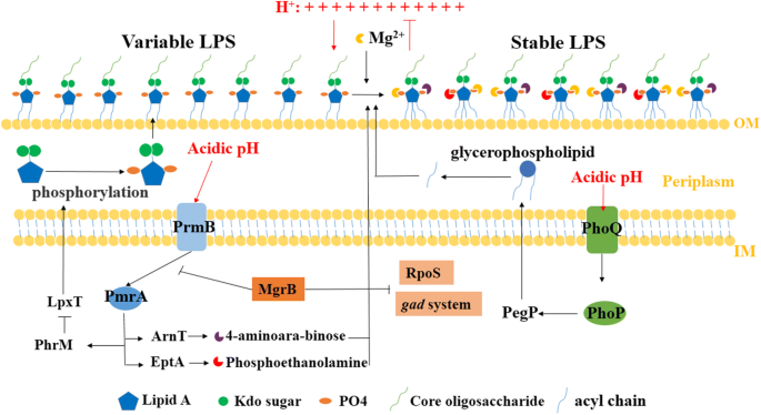


The Role Of Bacterial Cell Envelope Structures In Acid Stress Resistance In E Coli Springerlink
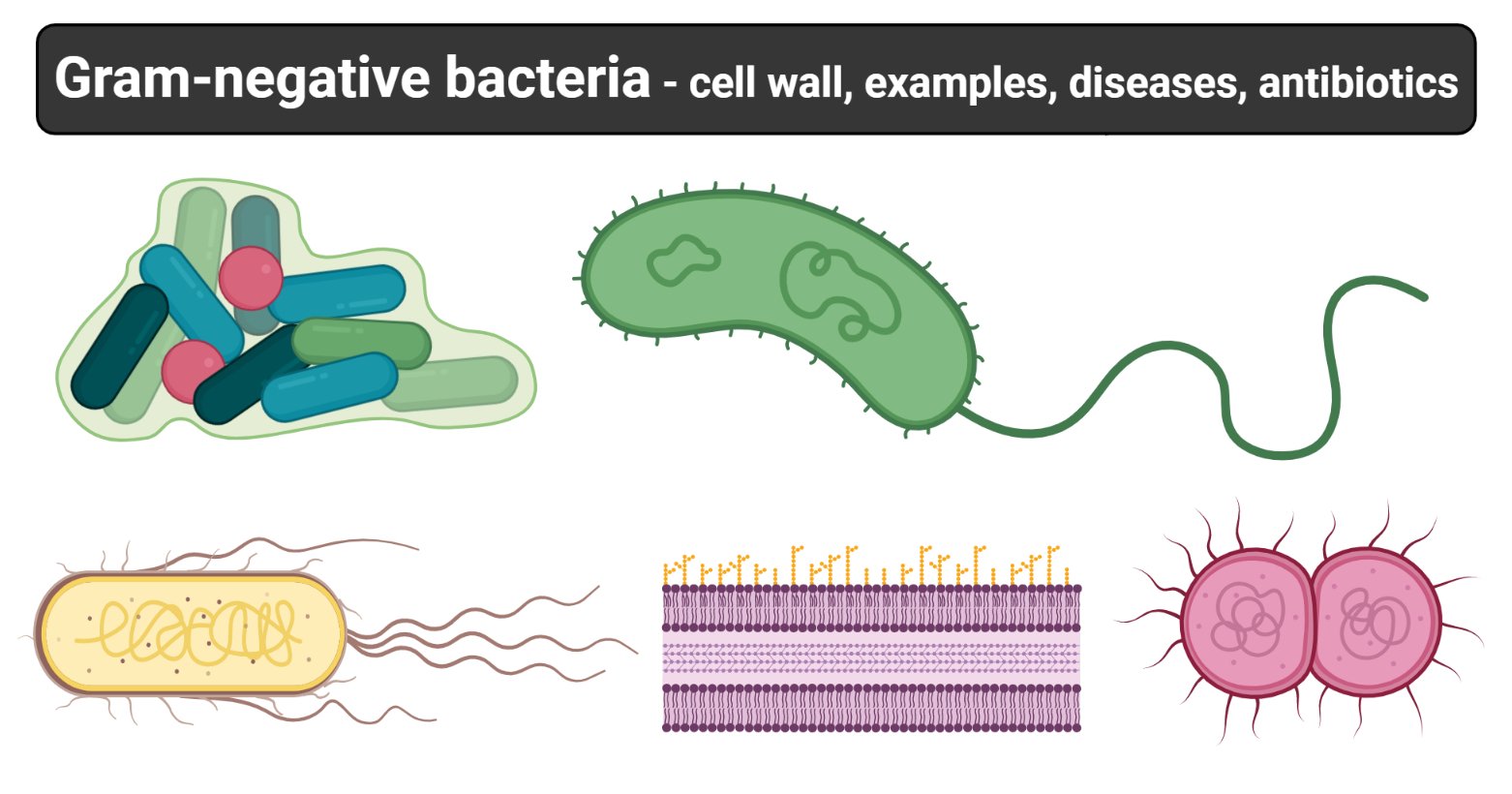
Gramnegative bacteria have a cell wall structure that is unable to retain the crystal violet dye Majority of the Gramnegative bacteria are pathogens owing to the characteristic outer membrane of the cell wall Escherichia coli is the most commonly known Gramnegative bacteriumIt is in fact an integral compartment of the gramnegative cell wall (5) Together the plasma membrane and the cell wall (outer membrane, peptidoglycan layer, and periplasm) constitute the gramnegative envelope (5, 9)Incorporation of new cell wall in differently shaped bacteria Rodshaped bacteria such as B subtilis or E coli have two modes of cell wall synthesis new peptidoglycan is inserted along a helical path (A), leading to elongation of the lateral wall, and is inserted in a closing ring


Diagram Demonstrating Of The Cell Wall Structure Of A Grampositive Download Scientific Diagram


Pin On Science
More specifically, the arrangement of PGN in the cell wall of E coli is such that numerous hydroxyl and amide groups on the sugar backbone and the peptide chains are available for hydrogen bonds (Figure 3A) In addition, there are three negatively charged carboxyl groups and one positively charged amine group on the noncrosslinked peptide chains (on residue Dglutamate, residue Dalanine, and mDAP) that can form salt bridges with TolR periplasmic domainE coliK30 CPS Group I Group II 8αNeu5Ac2 9αNeu5Ac2 n E coliK92 CPS 3βDRib1 7βKdo2 n E coliK23 CPS (Whitfield(Whitfield a and Valvano 1nd Valvano 1993 Bio993 Biosyntsynthesiesiss and expression and expression of cellsurface polysaccharides in gramnegative bacteria Adv Microbial Physiol )Gramnegative bacteria have a tripartite cell envelope with the cytoplasmic membrane (CM), a stressbearing peptidoglycan (PG) layer, and the asymmetric outer membrane (OM) containing lipopolysaccharide (LPS) in the outer leaflet


Lipoprotein Lpp Regulates The Mechanical Properties Of The E Coli Cell Envelope Nature Communications
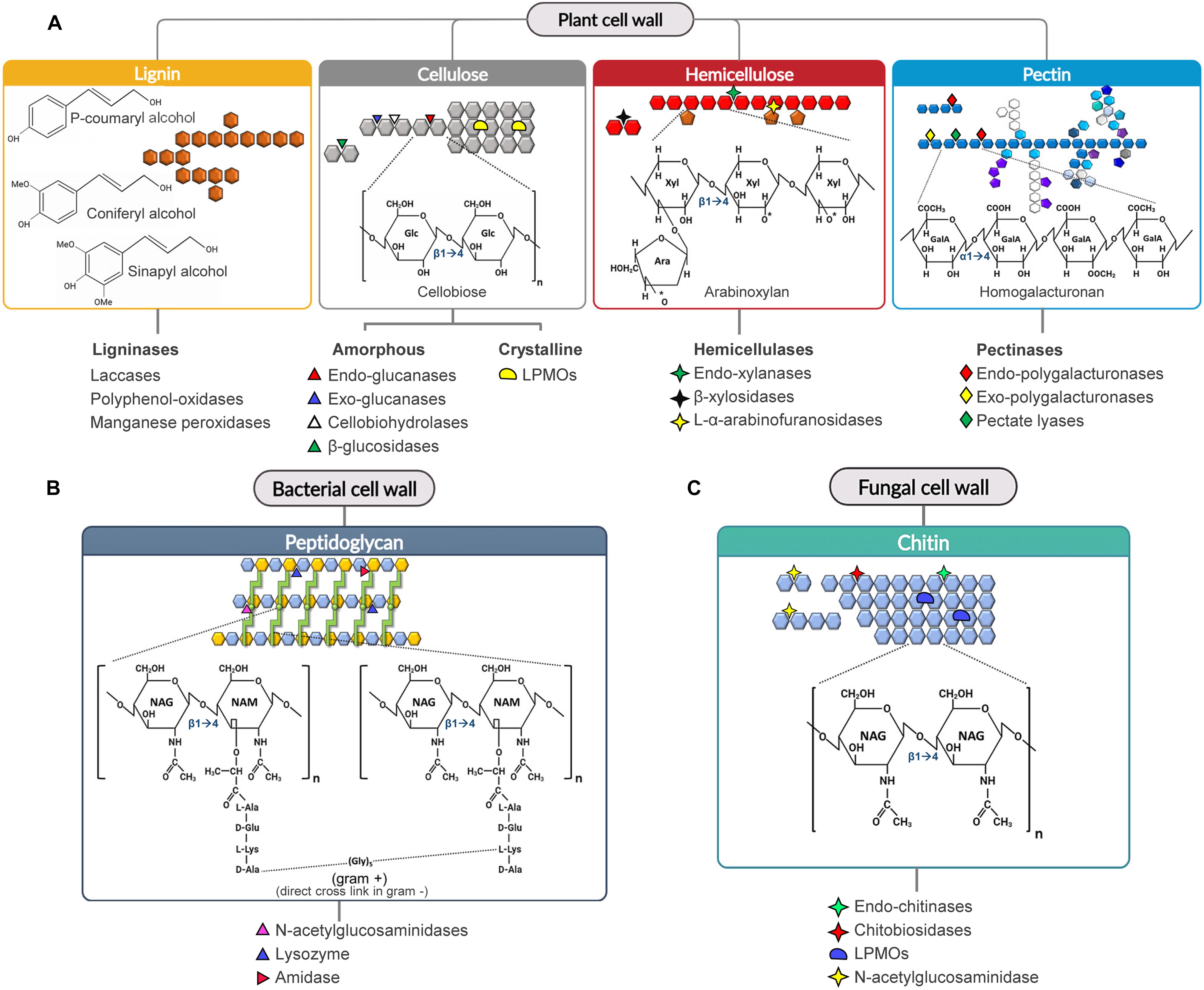


Frontiers Industrial Use Of Cell Wall Degrading Enzymes The Fine Line Between Production Strategy And Economic Feasibility Bioengineering And Biotechnology
Flagellar Hantigen of E coli contains protein flagellin, which is heatsensitive and can be destroyed at a temperature above 56°С Capsular, or Kantigen is composed of complex polysaccharides It covers cell wall Oantigen and preserves it against the actions of phagocytes and antibodiesThe cell wall structure of a bacterium decides the Gram character of the bacteria Grampositive bacteria have cell walls comprising a rich mesh of peptidoglycan layers that enable them to retain the dye Gramnegative bacteria, on the other hand, have a very thin peptidoglycan layer, and hence are unable to trap the dye moleculesThe bacterial cell wall plays a crucial role in viability and is an important drug target In Escherichia coli, the peptidoglycan crosslinking reaction to form the cell wall is primarily carried out by penicillinbinding proteins that catalyse D,Dtranspeptidase activity However, an alternate crosslinking mechanism involving the L,Dtranspeptidase YcbB can lead to bypass of D,Dtranspeptidation and betalactam resistance


Bacterial Outer Membrane Wikipedia
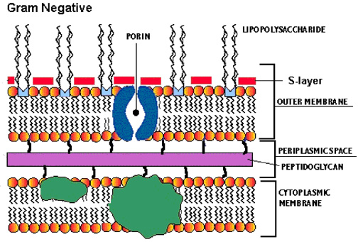


Proteobacteria Microbewiki
Non Acid Fast Bacteria Escherichia coli is an example of acid fast bacteria Conclusion Acid fast and non acid fast bacteria are two types of bacteria, which can be differentiated based on the presence of mycolic acid in the cell wall of the bacteria Acidfast staining is the technique used in discriminating the two types of bacteriaShapes are not directly dependent on the chemical composition of the structure in question ()For instance, E coli cells treated with penicillin can have the same shape as glass tubes manipulated by a glass blower (reference118 and references therein)The cell wall of the bacteria is made up mostly of one large molecule called peptidoglycan Researchers from Hyderabad have identified an enzyme that plays a crucial role in the enlargement and


Structure And Function Of Bacterial Cells


Www Bellarmine Edu Faculty Dobbins Secret readings Lecture notes 313 Ch03 Pdf
1 Describe peptidoglycan structure 2 Compare and contrast the cell walls of typical Grampositive and Gramnegative bacteria 3 Relate bacterial cell wall structure to the Gramstaining reaction 37The fimbriae are of type 1 (hemagglutinating & mannosesensitive) and are present in both motile and nonmotile strains Some strains of E coli isolated from extraintestinal infections have a polysaccharide capsule They are nonsporing They have a thin cell wall with only 1 or 2 layers of peptidoglycanThe Tphages, E coli B serves as a host l The receptor for the T3 and T4 and T7 phages was shown to be the cell wall lipopolysaccharide l 41 However, the structure of the E coli B lipopolysaccharide has hitherto remained unknown For these reasons and in conjunction with our studies on E coli R lipopoly


Plasticity Of Escherichia Coli Cell Wall Metabolism Promotes Fitness And Antibiotic Resistance Across Environmental Conditions Elife


Phospholipid Distribution In The Cytoplasmic Membrane Of Gram Negative Bacteria Is Highly Asymmetric Dynamic And Cell Shape Dependent Science Advances
MORPHOLOGY OF ESCHERICHIA COLI (E COLI) Shape – Escherichia coli is a straight, rod shape (bacillus) bacterium Size – The size of Escherichia coli is about 1–3 µm × 04–07 µm (micrometer) Arrangement Of Cells – Escherichia coli is arranged singly or in pairs Motility – Escherichia coli is a motile bacterium Some strains of E coli are nonmotileStructurally, a gramnegative cell wall consists of two layers external to the cell membrane a thin layer of peptidoglycan (too thin to absorb a significant amount of methyl violet stain), and an outer membrane (unique to gramnegative bacteria) that typically contains porins that facilitate the diffusion of small (Cell rigidity was achieved by thickening the cell walls via insertion of a constitutive gltA (encoding citrate synthase) promoter in front of a series of cell wall synthesis genes on the chromosome of several E coli derivatives, resulting in 132–160 folds increase of Young's modulus in mechanical strength for longer E coli cells overexpressing fission ring FtsZ protein inhibiting gene sulA


Edta Weakens The E Coli Cell Envelope A Length Of The Cell Wall Download Scientific Diagram


Team Siat Scie Design 18 Igem Org
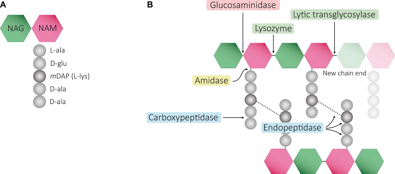
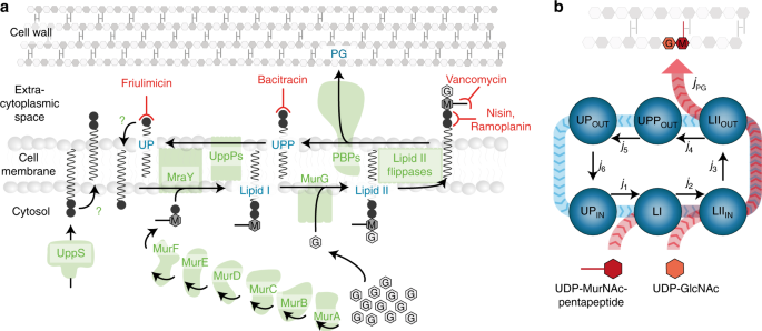
The cell wall of bacteria is composed of interlinked chains of peptidoglycan which both;Cell structure, metabolism & life cycle E coli serotype O157H7 is a mesophilic, Gramnegative rodshaped (Bacilli) bacterium, which possesses adhesive fimbriae and a cell wall that consists of an outer membrane containing lipopolysaccharides, a periplasmic space with a peptidoglycan layer, and an inner, cytoplasmic membraneBacterial cells are typically surrounded by an essential netlike macromolecule called the cell wall This structure is constructed of peptidoglycan (PG), a unique bacterial heteropolymer consisting of glycan chains of Nacetylmuramic acid (MurNAc) and Nacetylglucosamine (GlcNAc) repeating units with attached stempeptides used to form the matrix crosslinks Many of our most effective antibiotic therapies target cell wall biogenesis, and much of what we know about the cell wall assembly


Q Tbn And9gctnd0bk6ls8yuqlxdj9rtdvvfq Q5uj8cr Vbuzljzqmzbhrtcb Usqp Cau


Bacterial Cell Structure Wikipedia
E coli growing on basic cultivation media E coli stains Gramnegative because its cell wall is composed of a thin peptidoglycan layer and an outer membrane During the staining process, E coli picks up the color of the counterstain safranin and stains pink(Braun, 1975) It is the only known protein in E coli to be covalently attached to the PGN of the cell wall It exists in two states 33% of the lipoprotein is covalently bound to the cell wall via a peptide bond, and 66% is free in the periplasm BLP is proposed to have a primarily structural function, essentially actingMurNAc is unique to bacterial cell walls, as is Dglu, DAP and Dala The muramic acid subunit of E coli is shown in Figure 16 below Figure 16 The structure of the muramic acid subunit of the peptidoglycan of Escherichia coli This is the type of murein found in most Gramnegative bacteria


Bacteria 101 Cell Walls Gram Staining Common Pathogens Tusom Pharmwiki


Cell Wall Structure Of E Coli And B Subtilis Semantic Scholar
The cell wall of bacteria is made up of peptidoglycans called murein Bacteria contain 70S ribosomes, and bacterial DNA is arranged in the nucleoid Some bacteria may contain flagella for their movement The basic shapes of bacteria are coccus, bacillus, and spirillumMaintain the shape of the bacteria and act as a barrier to protect the bacteria from their often harsh environments•Cellassociated heteropolysaccharides •Most often acidic, but some are neutral •Solutions can be highly viscous •Grampositive and Gramnegative products •Lipopolysaccharides (LPSs) •Component of the outer membrane •Amphiphilic •Gramnegative bacterial component •Teichoic acid •GramPositive •Mycobacterium cell wall


Archaea Vs Bacteria Biology For Majors Ii


Bacteria 101 Cell Walls Gram Staining Common Pathogens Tusom Pharmwiki
Therefore, ProP also plays an important role in the structure of membrane wall in E coli The concentration of E coli cell bipolar permeability transporter ProP increases with the increase of cardiolipin content and depends on the structure of the cytoplasmic carboxyl terminal domain of the transporter 42Betweenthe cell wall structure ofS typhimurium and E coli strains and their resistance to the bacteriocidal lysosomal fraction of polymorphonuclear (PMN) leukocytes (1) havebeen demonstrated (6, 7) To obtain more detailed information on the influence of bacterial cell wall composition in ingestion and survival within the phagocyte, weThe structure of peptidoglycans of Escherichia coli and Bacillus subtilis is similar except for a few minor modifications, but murein (cell wall) structures are extremely different because the major cell wall constituents, anionic polymers, are not attached to peptidoglycans of E coli but are attached to those of B subtilis


Murein Peptidoglycan Structure Architecture And Biosynthesis In Escherichia Coli Sciencedirect


Peptidoglycan Chain Synthesis Of The E Coli Cell Wall Download Scientific Diagram
1 Describe peptidoglycan structure 2 Compare and contrast the cell walls of typical Grampositive and Gramnegative bacteria 3 Relate bacterial cell wall structure to the Gramstaining reaction 37Gramnegative bacteria such as Escherichia coli are surrounded by an outer membrane, which encloses a peptidoglycan layer Even if thinner than in many Grampositive bacteria, the peptidoglycan in E coli allows cells to withstand turgor pressure in hypotonic medium In hypertonic medium, E coli treated with a cell wall synthesis inhibitor such as penicillin G form walldeficient cellsE coli stains Gramnegative because its cell wall is composed of a thin peptidoglycan layer and an outer membrane During the staining process, E coli picks up the color of the counterstain safranin and stains pink The outer membrane surrounding the cell wall provides a barrier to certain antibiotics such that E coli is not damaged by penicillin


3


Q Tbn And9gcqoeecbggqeoibpsy9spzi9ifn Utadjplkmtyyfwfcp3h6qdph Usqp Cau


Prokaryotic Cell Anatomy


A Structure Of The E Coli Bacterial Cell Envelope Showing The Outer Download Scientific Diagram


Regulation Of Bacterial Cell Wall Growth Egan 17 The Febs Journal Wiley Online Library
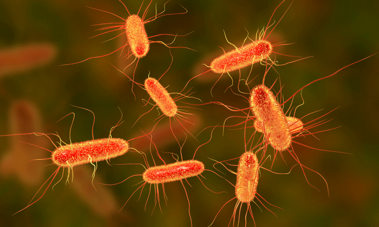


Bacteria Outer Membranes May Be Key To New Antibiotics
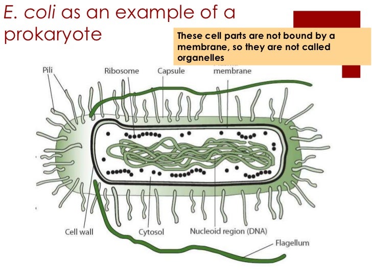


2 2 Prokaryotic Cells


Elucidating Peptidoglycan Structure An Analytical Toolset Trends In Microbiology


E Coli Cell Wall Structure Page 1 Line 17qq Com


Structure And Function Of Bacterial Cells


Cell Shape And Cell Wall Organization In Gram Negative Bacteria Pnas
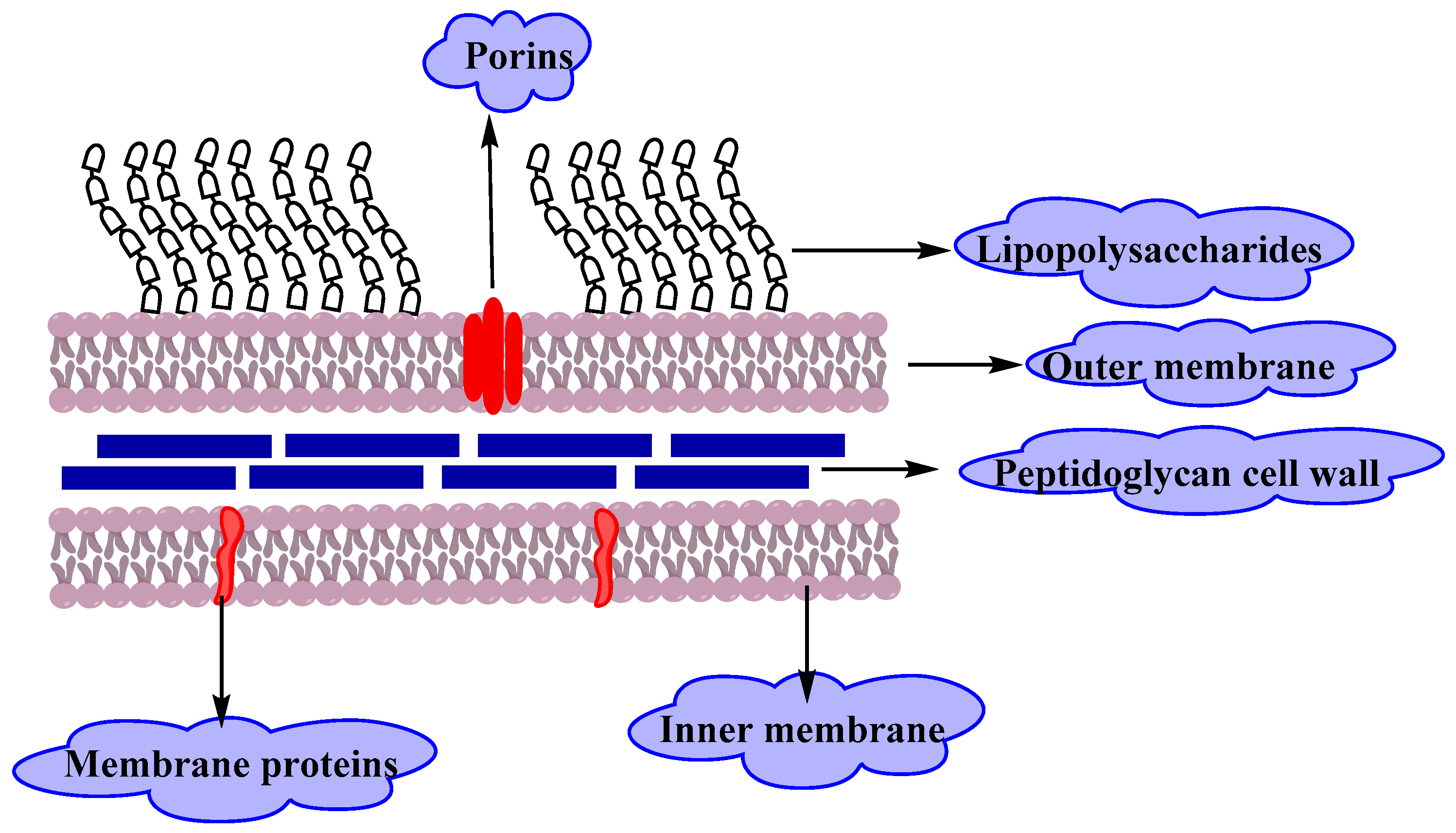


Molecules Free Full Text Resistance Of Gram Negative Bacteria To Current Antibacterial Agents And Approaches To Resolve It Html


Q Tbn And9gcqksoeyhk0r7phnhaimry3xpw1noc8z1ifj7d7yysogp61j 26c Usqp Cau


Bacterial Cell Wall An Overview Sciencedirect Topics
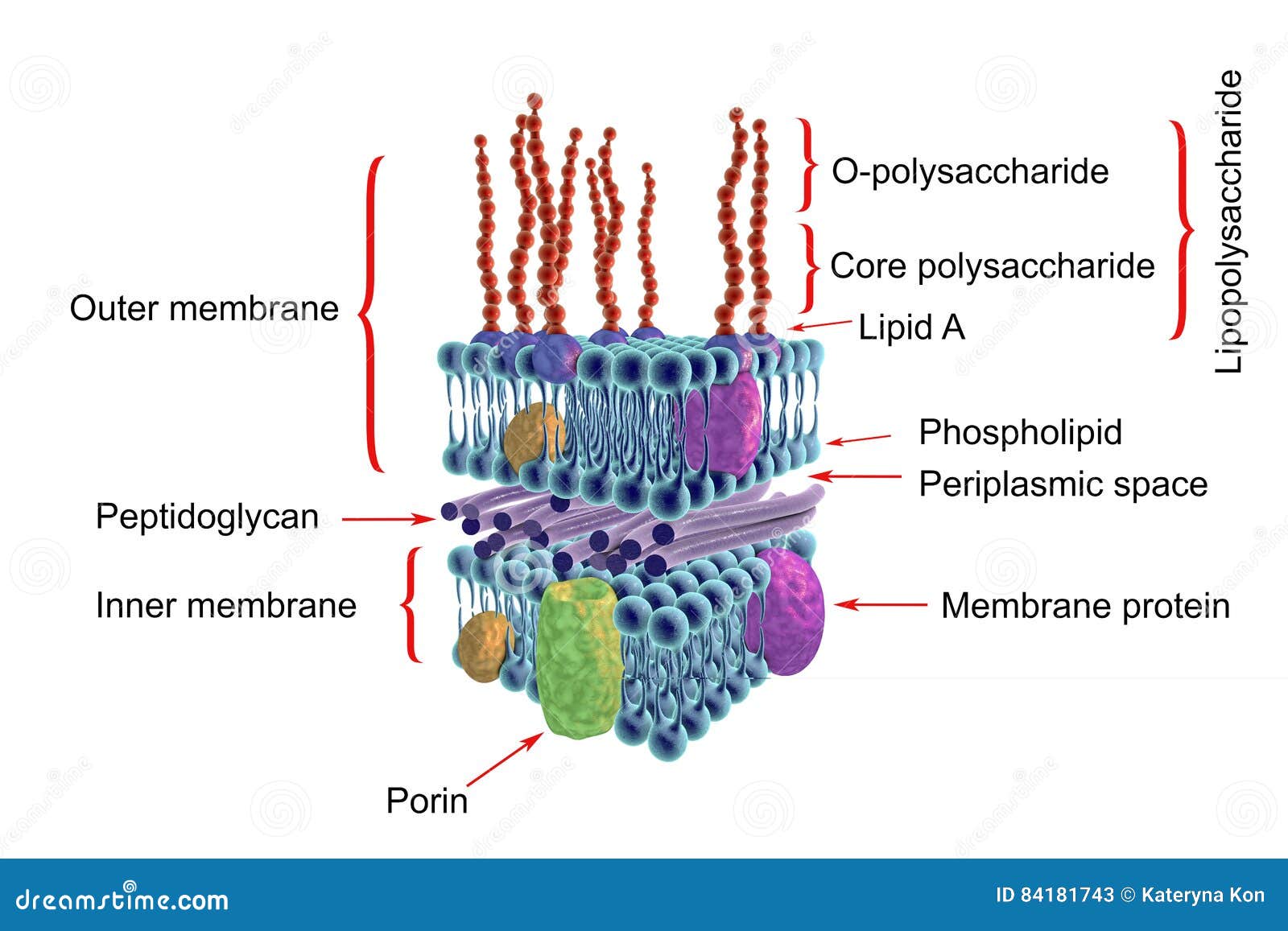


Structure Of Gram Negative Bacteria Cell Wall Stock Illustration Illustration Of Lipopolysaccharide Plasma


Chapter 2 Cells


Bioactive Cell Like Hybrids From Dendrimersomes With A Human Cell Membrane And Its Components Pnas


Prokaryotic Cells Bioninja
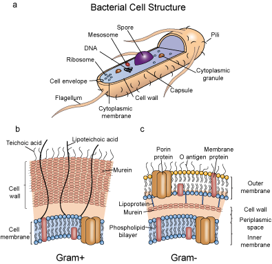


Bacterial Antigens Creative Diagnostics


Peptidoglycan Hydrolase Of An Unusual Cross Link Cleavage Specificity Contributes To Bacterial Cell Wall Synthesis Pnas


Bacterial Membrane An Overview Sciencedirect Topics
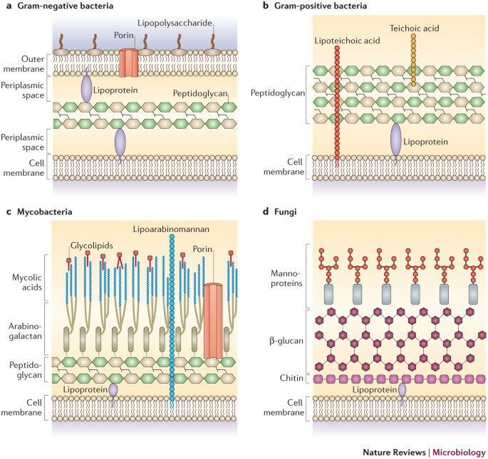


Through The Wall Extracellular Vesicles In Gram Positive Bacteria Mycobacteria And Fungi Nature Reviews Microbiology


The Bacterial Outer Membrane Is An Evolving Antibiotic Barrier Pnas


Structure And Function Of Bacterial Cells


Dissecting Escherichia Coli Outer Membrane Biogenesis Using Differential Proteomics


Various Facets Of Pathogenic Lipids In Infectious Diseases Exploring Virulent Lipid Host Interactome And Their Druggability Springerlink


Bacterial Cell Wall Structure Composition And Types Online Biology Notes


Chapter1 Cell Structure Of Bacteria


4 Bacteria Cell Walls Biology Libretexts


Crystal Structure Of An Intramembranal Phosphatase Central To Bacterial Cell Wall Peptidoglycan Biosynthesis And Lipid Recycling Nature Communications


Frontiers Peptidoglycan Muropeptides Release Perception And Functions As Signaling Molecules Microbiology


Cell Wall Wikipedia


Antibacterial Activities Of Ag Ions On The E Coli Cell Wall Download Table


Plasticity Of Escherichia Coli Cell Wall Metabolism Promotes Fitness And Antibiotic Resistance Across Environmental Conditions Elife


Cell Wall Structures Of Gram Positive And Gram Negative Bacteria And Download Scientific Diagram


Antibacterial Mechanism Of Bacteriolyses Of Bacterial Cell Walls By Zinc Ion Induced Activations Of Pgn Autolysins And Dna Damages Scitechnol


Bioactive Cell Like Hybrids From Dendrimersomes With A Human Cell Membrane And Its Components Pnas


Bacterial Growth And Cell Division A Mycobacterial Perspective Microbiology And Molecular Biology Reviews


Phospholipid Distribution In The Cytoplasmic Membrane Of Gram Negative Bacteria Is Highly Asymmetric Dynamic And Cell Shape Dependent Science Advances


Bacterial Inclusion Bodies A Treasure Trove Of Bioactive Proteins Trends In Biotechnology


Regulation Of Bacterial Cell Wall Growth Egan 17 The Febs Journal Wiley Online Library


Enzyme Explorer Key Resources Lysing Enzymes Sigma Aldrich


Frontiers Cracking Open Bacterial Membrane Vesicles Microbiology


Structure And Function Of Bacterial Cells


Gram Negative Bacteria Cell Wall Examples Diseases Antibiotics


Unique Characteristics Of Prokaryotic Cells Microbiology


Escherichia Coli Adaptation And Response To Exposure To Heavy Atmospheric Pollution Scientific Reports


Bacteria Cell Walls General Microbiology


Bacteria 101 Cell Walls Gram Staining Common Pathogens Tusom Pharmwiki


Minimal Exposure Of Lipid Ii Cycle Intermediates Triggers Cell Wall Antibiotic Resistance Nature Communications


4 Bacteria Cell Walls Biology Libretexts


Molecular Mechanisms Of Membrane Targeting Antibiotics Sciencedirect


Pin On Photoshop Png


Structure Of Lipopolysaccharide Lps Lps Is Found On The Cell Wall Of Download Scientific Diagram
:max_bytes(150000):strip_icc()/gram_negative_bacteria-5b7f308646e0fb002cbcdc5d.jpg)


Gram Positive Vs Gram Negative Bacteria


Cell Wall Structures Of Gram Positive And Gram Negative Bacteria And Download Scientific Diagram


Increasing The Permeability Of Escherichia Coli Using Mac Scientific Reports


Impact Of Membrane Phospholipid Alterations In Escherichia Coli On Cellular Function And Bacterial Stress Adaptation Journal Of Bacteriology


Cell Wall Structure Of E Coli And B Subtilis Semantic Scholar


E Coli Cell Structure Page 1 Line 17qq Com


Frontiers Maintaining Integrity Under Stress Envelope Stress Response Regulation Of Pathogenesis In Gram Negative Bacteria Cellular And Infection Microbiology


2 2 Prokaryotic Cells Bioninja


Pin On Study


Bacteriolyses Of Bacterial Cell Walls By Cu Ii And Zn Ii Ions Based On Antibacterial Results Of Dilution Medium Method And Halo Antibacterial Test
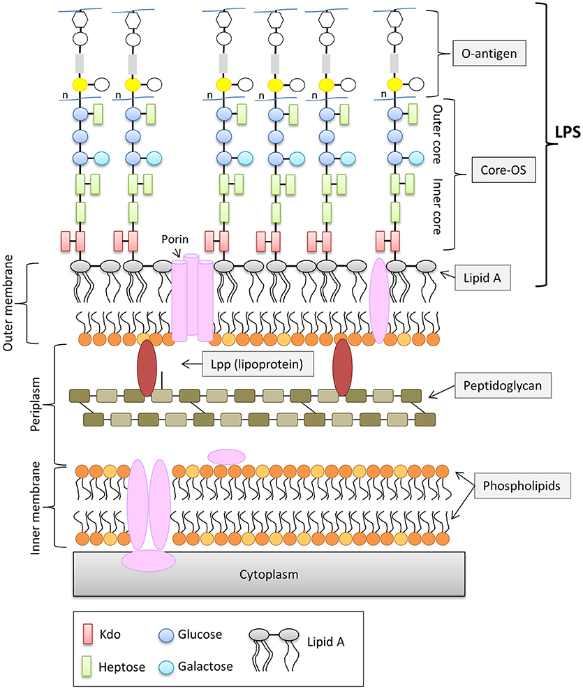


Frontiers The Role Of Outer Membrane Proteins And Lipopolysaccharides For The Sensitivity Of Escherichia Coli To Antimicrobial Peptides Microbiology
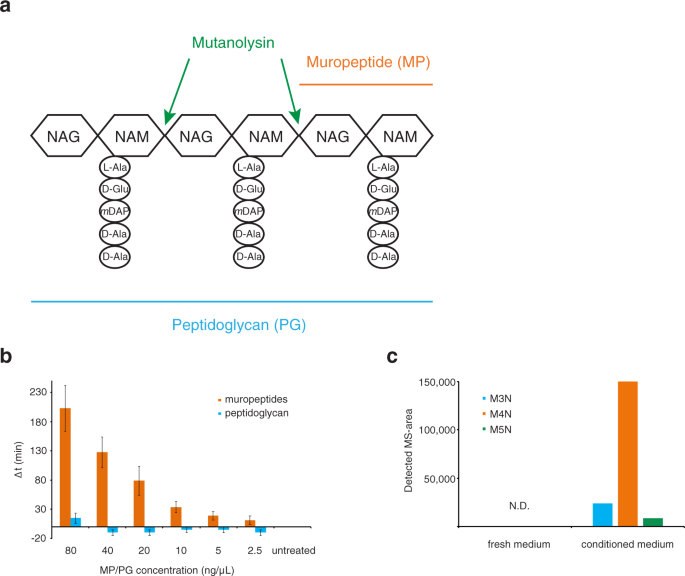


Muropeptides Stimulate Growth Resumption From Stationary Phase In Escherichia Coli Scientific Reports


Glycopedia


Converting Escherichia Coli Into An Archaebacterium With A Hybrid Heterochiral Membrane Pnas


Cell Wall Bacteria
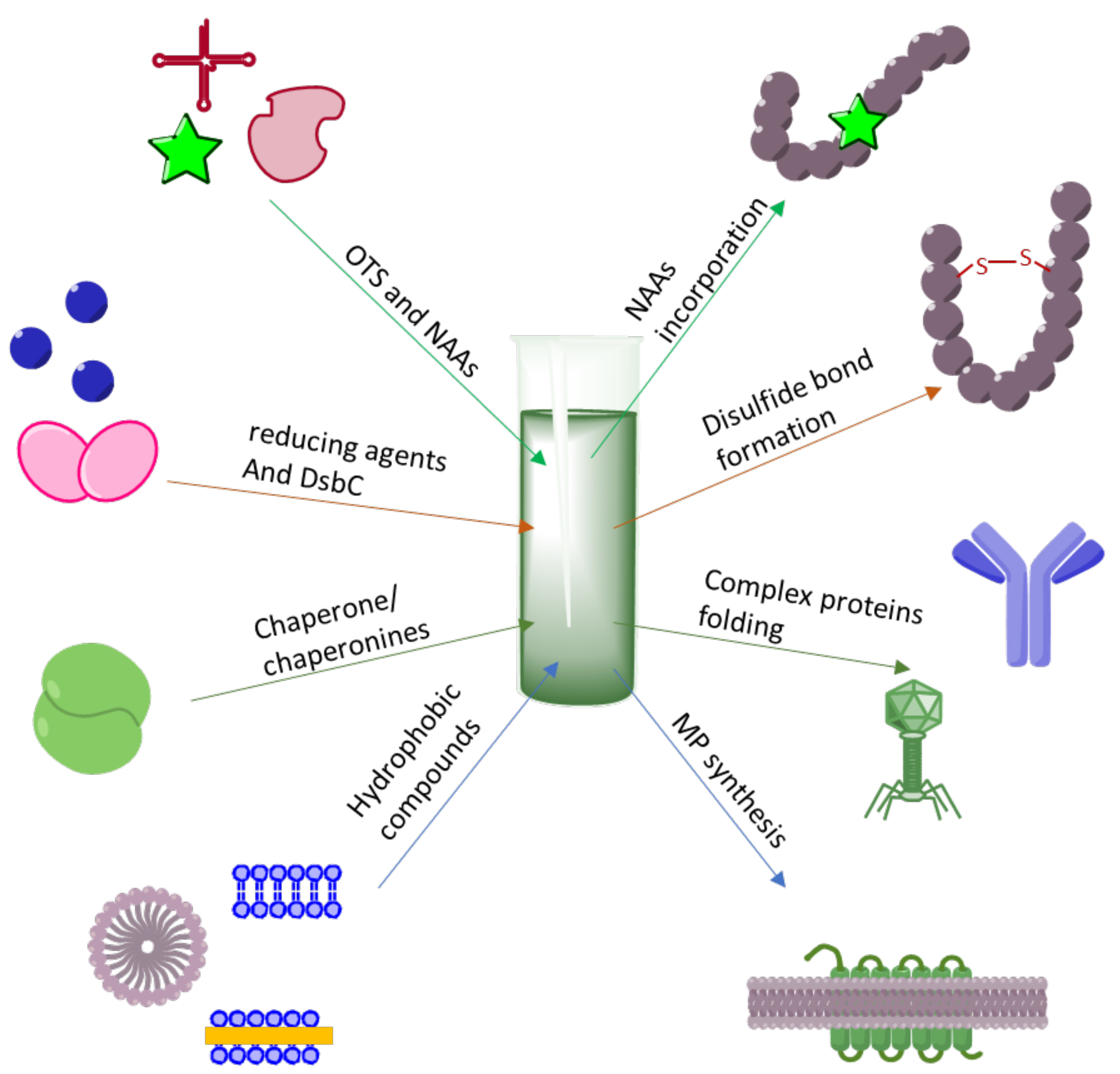


Ijms Free Full Text Escherichia Coli Extract Based Cell Free Expression System As An Alternative For Difficult To Obtain Protein Biosynthesis Html


Coordination Of Peptidoglycan Synthesis And Outer Membrane Constriction During Escherichia Coli Cell Division Elife
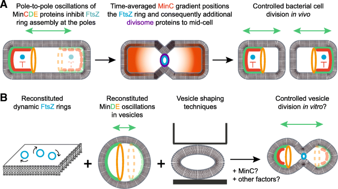


Synthetic Cell Division Via Membrane Transforming Molecular Assemblies Bmc Biology Full Text
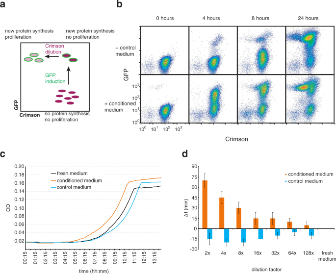


Muropeptides Stimulate Growth Resumption From Stationary Phase In Escherichia Coli Scientific Reports
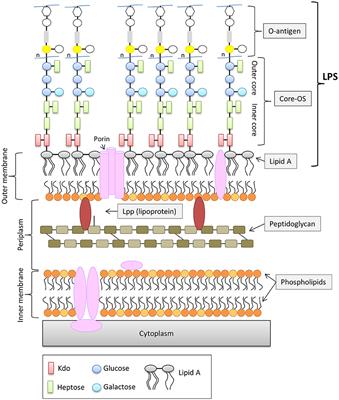


Frontiers The Role Of Outer Membrane Proteins And Lipopolysaccharides For The Sensitivity Of Escherichia Coli To Antimicrobial Peptides Microbiology


Gram Positive Bacterial Cell Envelopes The Impact On The Activity Of Antimicrobial Peptides Sciencedirect


Figure 1 From Advances In Understanding Bacterial Outer Membrane Biogenesis Semantic Scholar
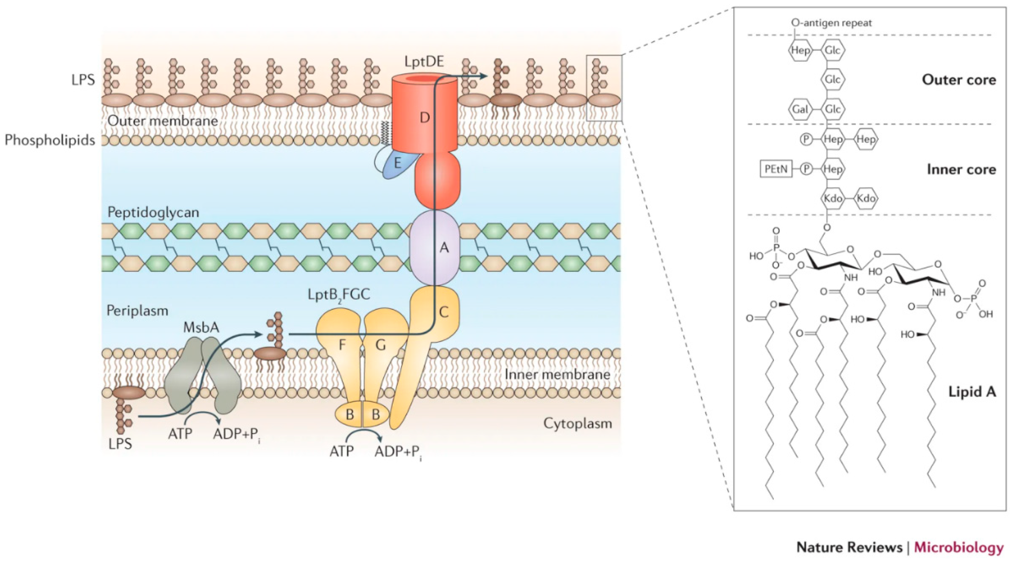


Biosensors Free Full Text Nanomaterials For Biosensing Lipopolysaccharide Html


Basic Biology Of Oral Microbes Sciencedirect


コメント
コメントを投稿